
By Dr Sen Le and Daniel Seo | Chief Clinical Officer & Chief Strategy Officer at Eyes of AI™
In our first article, we made a simple argument: dentistry’s next leap is not digital, but intelligence. Digital changed how we capture information. Intelligence changes what we do with it. Our images have never been sharper, nor our workflows faster. Yet our diagnoses are no more accurate than before.
This article is about why. The answer is uncomfortable. Modern imaging now holds far more information than the human eye was ever built to read.
We have spent two decades making the image sharper.
We have never asked whether the eye could keep up.
The image has outgrown the eye
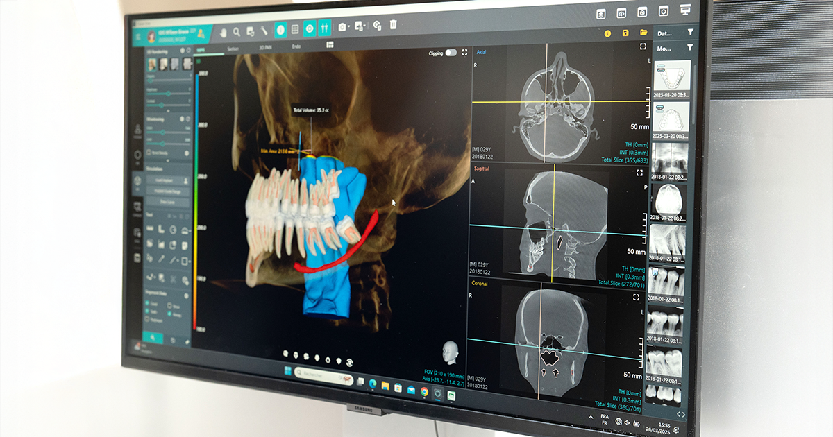
Modern imaging is staggering in its detail. A single bitewing typically holds two to four million pixels. A panoramic image can hold up to six million and every one of those pixels can encode more than 65,000 shades of grey. A cone-beam CT (CBCT) scan dwarfs them both with more than 1 billion voxels, each carrying thousands of shades of its own, up to 65,536 at the highest bit depths.1
The human eye can distinguish about 60 shades of grey.2
That is the gap. We now capture more than any eye can take in, then ask ourselves to find what matters inside it, often in seconds. A bitewing is read in under a minute.3 A CBCT that deserves twenty minutes may get less than five.4
And volume is only part of the difficulty. A flat radiograph collapses three-dimensional anatomy onto a single plane, so overlapping structures can hide pathology. The earliest and most treatable signs, such as incipient caries or subtle early bone loss, are faint, low in contrast, and easily lost.5,6 None of this is a failure of skill. It is the reality of human perception applied to a task that has quietly outgrown it.
What the evidence shows
The consequences are measurable. Reading radiographs unaided, dentists catch fewer than half of the early caries, the very lesions still small enough to reverse.5 Ask two skilled clinicians to assess the same images and they frequently disagree. In one reliability study, 14 dentists varied substantially in how they identified periapical lesions, and even the same clinician sometimes classified the same image differently when reviewing on a different occasion.7 This is not a question of skill. It is what happens to even the most expert eye, working at the very edge of what human perception allows.
Key insight: Variability in radiographic interpretation is not a gap in competence. It is the predictable result of asking expert clinicians to detect subtle disease across dense, greyscale images, at speed, and to do so consistently, image after image, patient after patient.
Periodontal disease: where the limits show
Periodontal disease is the clearest example. Despite being largely a preventable disease, it affects up to 50% of the global population with 1 billion people affected by its severe form. Due to its gradual and silent nature, it is no surprise that the average time from onset of disease to diagnosis has been found to be 56 months. Detecting it early means comparing fine changes across a full series, and ideally against images taken months or years earlier. That is exactly the slow, comparative pattern the eye handles poorly.
What augmentation changes
This is the practical case for intelligent systems, and it is narrower and more useful than the idea of AI replacing dentists. Think of it as spell-check for radiographs, not autopilot: it flags what you might have missed.
An AI system does not tire. It gives the last image of the day the same attention as the first, and it can compare bone levels across a full series, and over time, with a consistency no clinician can sustain across a busy week.
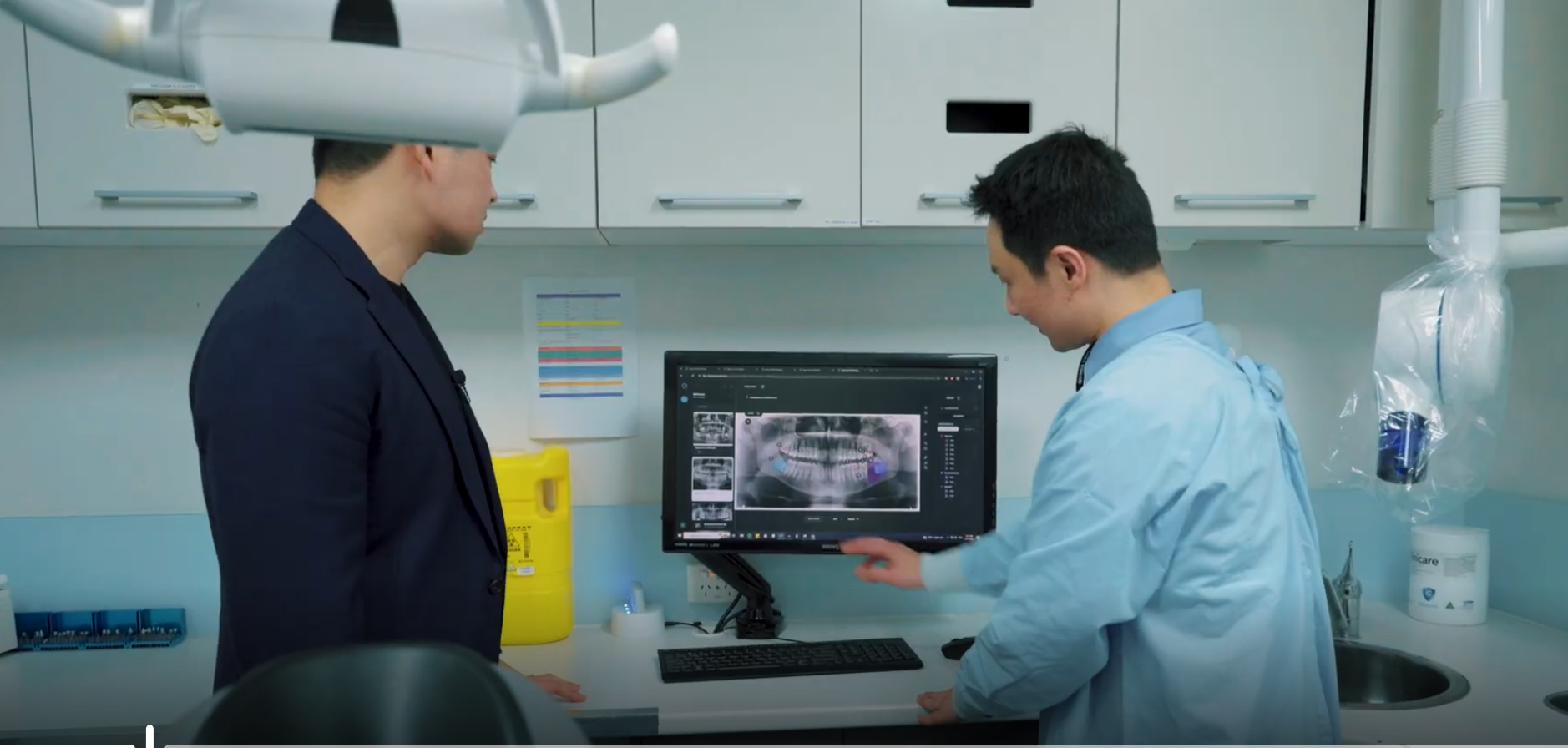
At Eyes of AITM, that is precisely our focus: to identify periodontal disease earlier, by surfacing the subtle bone-level changes that are easy to overlook, and to screen more efficiently with less variability, by giving every image the same structured review. The clinician still decides. The system simply reduces the noise around that decision.
For your practice: Augmented screening is most valuable where manual reading is weakest: subtle, slow-moving, multi-site conditions such as periodontal disease. A consistent second read helps catch early findings and standardise interpretation across your team.
Reframing the problem
For too long, we have treated diagnostic variability as a problem of effort: more training, more care, more time. These help. But they cannot close a gap this wide. The question was never how hard we look. It is that we have been asking human eyes to do what no eye was built to do.
Recognising that is not a criticism of the profession. It is the first honest step toward a dentistry that matches the images we now produce.
We have spent twenty years making the image sharper. The task now is to make it easier to understand, so that less disease is missed, and no clinician has to shoulder that burden alone.
In our next article, we turn to the data behind those images: the vast clinical record dentistry generates every day, and how little of it we currently use.
References
- Farman AG, Farman TT. A comparison of 18 different X-ray detectors currently used in dentistry. Oral Surg Oral Med Oral Pathol Oral Radiol Endod. 2005;99(4):485-9. doi:10.1016/j.tripleo.2004.04.002.
- Queiroz PM, Fardim KC, Costa AL, Matheus RA, Lopes SL. Texture analysis in cone-beam computed tomographic images of medication-related osteonecrosis of the jaw. Imaging Sci Dent. 2023;53(2):109-115. doi:10.5624/isd.20220202.
- Patel HB, Marwaha J, Hirani N, Suthar P, Arya A, Pandey N. Diagnostic accuracy and efficiency of AI-assisted radiographic interpretation compared to conventional methods in early detection of dental caries. J Pharm Bioallied Sci. 2025;17(Suppl 4):S3192-S3194. doi:10.4103/jpbs.jpbs_1278_25.
- Ezhov M, Gusarev M, Golitsyna M, Yates JM, Kushnerev E, Tamimi D, et al. Clinically applicable artificial intelligence system for dental diagnosis with CBCT. Sci Rep. 2021;11(1):15006. doi:10.1038/s41598-021-94093-9.
- Devlin H, Williams T, Graham J, Ashley M. The ADEPT study: a comparative study of dentists’ ability to detect enamel-only proximal caries in bitewing radiographs with and without the use of AssistDent artificial intelligence software. Br Dent J. 2021;231(8):481-5. doi:10.1038/s41415-021-3526-6.
- Krois J, Ekert T, Meinhold L, Golla T, Kharbot B, Wittemeier A, et al. Deep learning for the radiographic detection of periodontal bone loss. Sci Rep. 2019;9(1):8495. doi:10.1038/s41598-019-44839-3.
- Meusburger T, Wülk A, Kessler A, Heck K, Hickel R, Dujic H, et al. The detection of dental pathologies on periapical radiographs—results from a reliability study. J Clin Med. 2023;12(6):2224. doi:10.3390/jcm12062224.
About Eyes of AI™
Eyes of AI™ is an Australian-founded MedTech company developing AI-powered tools to transform dental diagnostics and
workflows. Its platform spans 2D and 3D imaging, including automated pathology detection, cephalometric analysis, and advanced CBCT segmentation. By combining clinical expertise with cutting-edge artificial intelligence, Eyes of AI enables faster, more accurate, and scalable decision-making for dentists. The company works closely with leading institutions such as CSIRO and the University of Sydney, and partners with global manufacturers to integrate AI directly into clinical workflows. Its mission is to improve patient outcomes and expand access to high-quality dental care through intelligent, practical technology.
Website: https://eyesofai.com/